
Shining New Light on Skin Cancer Detection
A research group in the UT Austin Biomedical Engineering Department is examining how to improve the efficacy of skin cancer detection and removal.
The current standard procedure, known as Mohs micrographic surgery, is considered quite accurate. However, it requires a full laboratory adjacent to the procedure room to determine if the physician successfully removed the full tumor.
The process involves cutting out tissue and using a microscope to determine if parts of the tumor remain in the body. Consequently, the physician needs to remove additional tissue to ensure the tumor is fully removed.
While highly accurate, the procedure is time-consuming. Each round of tissue removal involves additional time for analysis and requires highly specific equipment and training. This poses logistical challenges for patients who are not in close proximity to major medical centers.
A new $2.7 million grant from the National Institutes of Health is supporting a collaborative project between the Cockrell School of Engineering and the Dell Medical School at The University of Texas at Austin to improve the Mohs procedure and make it more accessible. Additionally, researchers aim to take this approach and implement it in other types of cancer surgery.
“Reducing the infrastructure burden of the current procedure could improve the patient experience and expand the patient population that has access to this highly accurate surgical approach,” said James Tunnell, professor of Biomedical Engineering.
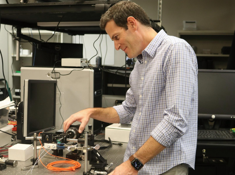
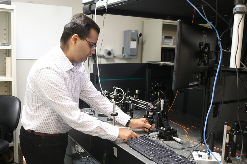
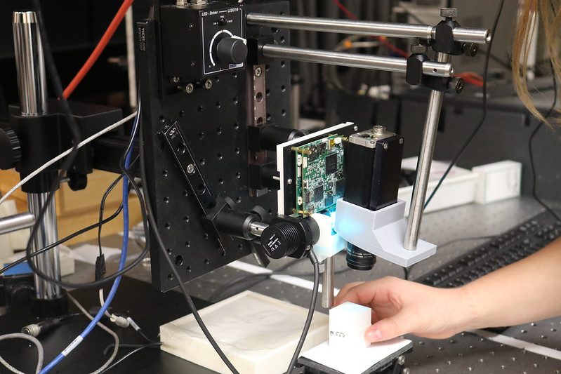
Tunnell’s team is working to create a box-shaped device that is equipped with cutting-edge microscopy tools and machine learning capabilities that can analyze a tissue sample in a matter of minutes.
The surgeon can determine if they removed the entirety of the tumor, or if another round of tissue removal is required. Mohs surgeons typically complete a year-long fellowship in micrographic surgery and dermatologic oncology, which includes significant time with histology—the study of tissue structure using microscopes.
“We look at the entire periphery and the base of what has been removed,” said Matthew Fox, a Mohs-certified surgeon, chief of the Division of Dermatology & Dermatologic Surgery, associate professor of the Department of Internal Medicine at Dell Med and one of the leaders of the project. “Think about a Reese’s Peanut Butter Cup; we are analyzing the wrapper that holds the chocolate and peanut butter.”
Each round of analysis takes time, and the process continues until there are no more tumor cells at the sample margin. This lengthy process occurs while the patient sits in an outpatient facility. The device that Tunnell’s team is developing would complete the analysis, making the process more efficient and potentially broader in accessibility.
Skin cancer is often readily accessible to a surgical team and can be removed with local anesthesia alone—this is what makes Mohs surgery the standard treatment protocol.
If the principles of Mohs surgery can be replicated with Tunnell’s device efficiently and simplified in the way the researchers envision, it could be applied to many other types of cancer, and its level of accuracy could be replicated elsewhere.
“More accurate identification of tumor margins during surgery may reduce additional surgeries and treatments,” Tunnell said.
Other research team members include biomedical engineering professor Mia Markey, associate professor of dermatology Tyler Hollmig, Dell Medical School dermatology professor Jason Reichenberg, and Brian Hobbs, associate professor of population health at Dell Medical School.
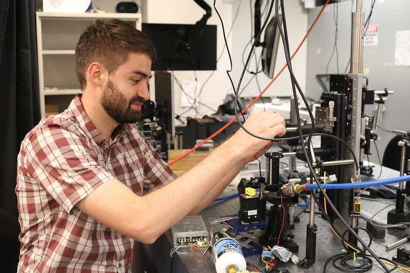
In addition to early cancer detection, Tunnell specializes in biomedical optical spectroscopy and imaging, laser-tissue interactions, nanotechnology, and nanophotonics. He is the principal investigator of the Biophotonics Laboratory at UT Austin and his research focuses on developing minimally invasive optical technologies for the diagnosis and treatment of disease.
It is widely believed that the greatest achievement that can be made in cancer management is the early detection and subsequent treatment of the disease. The next generation cancer management strategies require technologies that combine sensing, targeting, and treating of the earliest-stage disease.
Tunnell’s approach combines optical imaging, spectroscopy, and nanotechnology to develop systems capable of combined diagnosis and treatment of early cancer. In addition, Tunnell’s lab actively studies the basic mechanisms of light-tissue interactions to understand light transport and develop novel imaging strategies.